Category Archives: photosynthesis

Superphotosynthesizer: Cat Island Baldcypress

Today May 18, 2015 is ‘Fascination of Plants Day,’ an initiative organized by the European Plant Science Organisation with other events organized by the American Society of Plant Biology. On this blog, there’s no shortage of reasons why plants are fascinating, but to most they are still just the scenery. Take some time today to consider all that these primary producers do for you. Here are just a few things plants do for us- food, forestry products, paper, pharmaceuticals, energy, and beauty.

Of course, I am partial to the oxygen that they provide for us. In that spirit, today’s post will feature another superphotosynthesizer: the Cat Island Baldcypress located on the Cat Island National Wildlife Refuge in West Feliciana Parish, LA. This tree is the national champion of its species and also noted as the largest tree of any species east of the Sierra Nevada range.

It is located at the end of an easy walking trail (0.75 mile round-trip), but it only accessible for part of the year. Access to portions of the Cat Island NWR is prevented by levels of the Mississippi River since at least a couple of low bridges must be traversed to get you from the main road to the trailhead. If the river stage at Baton Rouge is greater than 20 feet, which is usually between February and June, there is no vehicle access to the trail. I was able to make a trip there in early February just before the river restricted access. It’s not quite clear whether the base of the tree itself is submerged at any point during the spring flooding because there is a really nice decking just before the tree at the end of the trail. As of today, the river stage at Baton Rouge is 27.8 feet, so it may still be another month before access is regained.

038

The tree is impressive. At 96 feet it is taller than all the other trees around, but it’s certainly not the tallest tree east of the Sierra Nevada. However, its girth is undeniably impressive. It has the characteristic buttressed-base of all baldcypress trees, which measures 17 feet in diameter and 56 feet in circumference.  It has knees as tall as me. Well, for those of you who know me in real life maybe that’s not so impressive, but for a random root outgrowth that is still significant.

037

This brings me to one of the real secrets of the swamp- cypress knees. These strange growths at the base of cypress trees have been puzzling botanists and plant biologists for centuries since Francois Andre Michaux wrote in 1819, “No cause can be assigned for their existence.” Many people have had theories as to how they contribute to cypress biology- increased aeration capability for growing in inundated swamps, methane (swamp gas) emission conduits, vegetative reproduction, mechanical support, nutrient acquisition, and carbohydrate storage. None of these hypotheses have really held up to analysis and the biological function* of these root outgrowths are still fascinating plant biologists today.

032

This is just one local fascinating plant example. Check out the links below for more information about Fascination of Plants Day or follow #FOPD on social media.

 029

Johnna

*These expendable appendages are painted and carved for folk art projects. They are also fairly proficient at disemboweling lawnmowers of homeowners with cypress trees in their yards and capsizing careless motorboat operators in the swamps. Perhaps this is a plant defense mechanism ahead of its time.

References and Links:

http://www.plantday.org/

http://fascinationofplantsday.org/home.htm

http://blog.aspb.org/fascination-of-plants-day/about-fascination-of-plants-day/

http://blog.aspb.org/fascination-of-plants-day/

http://www.fws.gov/refuges/profiles/index.cfm?id=43697

http://www.fws.gov/refuge/Cat_Island/visit/plan_your_visit.html

http://www.monumentaltrees.com/en/trees/baldcypress/deep_south/

http://www.na.fs.fed.us/pubs/silvics_manual/Volume_1/taxodium/distichum.htm

http://en.wikipedia.org/wiki/Cypress_knee

http://www.venerabletrees.org/good-knees-said-baldcypress/

http://arnoldia.arboretum.harvard.edu/pdf/articles/2000-60-4-cypress-knees-an-enduring-enigma.pdf

On becoming a tree…

“As long as you keep getting born, it’s all right to die sometimes”

Orson Scott Card, The Speaker for the Dead.

I’ve written many times about the differences between autotrophs and heterotrophs on this blog. Loyal readers will know I have a hard-line stance that humans fall decidedly into the heterotroph camp. Nevertheless, some art projects blur the lines between person and plant. So I was more than a little intrigued when a link for the Bios Urn came across my Facebook feed.

BiosUrn

The product offers an alternative to the traditional cemetery as your eternal destination. It’s a special biodegradable urn in which your ashes (or a loved one’s or a pet’s ashes) can be placed along with a tree seed. The whole thing is planted in the ground at a cemetery or other special location. The recycled carbon atoms of your body become the growth medium for a tree that will grow and live on after your death.

biosurn2

It’s beautiful. I get it and as someone who often quotes the Lorax, I’m all for any excuse to plant trees. But honestly, the first thing it made me think of was my all-time favorite book The Speaker for the Dead by Orson Scott Card.* Within the world of this book, humans live on a colony planet that happens to have another species of sentient beings called the Pequeninos. These organisms spend the majority of their life cycle as mammal-like beings, but can pass on into a third life as a tree provided they have been vivisected. It’s pretty gruesome and obviously humans don’t work this way, so cultural misunderstandings ensue.

However, because I’m a stickler for scientific accuracy when it comes to plant science (Hey, someone’s gotta be.), I have one problem with the overly simplistic marketing scheme of the Bios Urn. You don’t actually become the tree. For those of you saying, “I know, I know I won’t be a tree, but in the circle of life my molecules will become part of this tree.” I’ll still be over here at my blog shaking my head, “No, that’s not really how that works.” Here’s a reminder of the photosynthetic equation:

6 CO2 + 6 H2O (+ light) → C6H12O6 + 6 O2

Plants accumulate carbon and mass from atmospheric CO2, not carbon ash or even organic carbon compounds in the soil. The truth is that if you planted your ashes with your tree seed, your carbon would still remain locked in the soil for many years until eventually it is metabolized by microorganisms in the soil and released as CO2 as part of their respiration. This is not a very quick way to turn over your carbon molecules, and your molecules have millennia before they ever become a part of any tree. If you truly wanted your carbon molecules to incorporate into some kind of plant matter, then you need to find a way to carbonate your deceased body. Human carbonated essence could be stored in canisters and then used to supply CO2 to your plant of choice. You could be part of that camellia bush in your front yard, your favorite LSU Oak tree, the General Sherman etc. Scientifically accurate, but good luck marketing that compared to cremation.**

All of this highlights a common misconception about photosynthesis- it’s hard to comprehend how mass can accumulate into living things from air. Our gut tells us mass must come from something more substantial. We memorize the photosynthetic equation in elementary school, but few of us grasp its consequences and really believe it.

“This is how humans are: We question all our beliefs, except for the ones that we really believe in, and those we never think to question.”

Orson Scott Card, The Speaker for the Dead

So, when you die, ask someone to plant a tree or something in your memory. Do it for someone else you’d like to remember, but ashes not really required. Or buy a BiosUrn or put the ashes in a degradable coir starter pot from your local garden center, but you’ll probably want to supplement them with some form of NPK fertilizer. It will help the seed more than your carbon atoms.

Johnna

*It’s a sequel to Ender’s Game for those of you that may not be SciFi freaks.

**Although, in-home soda fountains are a thing now, so if there is a way to carbonate deceased relatives and store them in the handy canisters… well, you get the idea.

Links and References:

https://urnabios.com/

http://en.wikipedia.org/wiki/Speaker_for_the_Dead

The Dangerous Double Life of a Distinctive Diazotroph

For many people around the world, tonight is one of the most anticipated nights of the year. In that spirit, here’s some new science, not under the sun, but under the moonlight and starlight. New research from Wegener, Nagarajan and Pakrasi* describe something new about the Photosystem II D1 protein, an unexpected source that photosynthesis researchers had long written off as conquered or fully explained.

Why would we care about photosynthesis at night anyway? It all comes down to the distinctive lifestyle of the cyanobacterium Cyanothece, which belongs to an interesting class called unicellular diazotrophs (word of the day). Like all cyanobacteria, they perform oxygenic photosynthesis but they have the bonus biochemical ability of nitrogen-fixation.

Nitrogen-fixers are capable of pulling nitrogen gas (N2) out of the air and turning it into ammonia which the cells can use as a nitrogen source for important molecules like protein and DNA. Other microbes can perform nitrogen-fixation, and you may remember them as the root nodule symbionts of legumes. If you’re not a nitrogen-fixer, you have to rely on ammonia or other nitrate compounds present in the soil, sea or your diet (depending on what kind of organism you are) to make the required nitrogenous biomolecules. Since the Earth’s atmosphere is 78% N2, you don’t have to be great at math to figure out that acquiring the ability to tap into the atmospheric nitrogen source advances your species in the game of life a considerable number of squares.

What does this have to do with photosynthesis? Why don’t more plants and cyanobacteria get in on this nitrogen-fixation biochemistry? There’s a critical fatal flaw in the photosynthesis/nitrogen-fixation biochemical tango. Nitrogenase, the enzyme responsible for the nitrogen-fixation reaction is hopelessly and irreversibly killed by oxygen. Oxygen destroys the protein enzyme that’s already been made and tells the genome not to even bother wasting energy trying to make it new. If you’ve been paying attention to the blog, oxygenic photosynthesizers like cyanobacteria and plants make oxygen via their PSII enzyme every time light shines on them. So for nonphotosynthetic soil bacteria and other microbes, it’s not a big deal to avoid oxygen and go anaerobic to fix nitrogen. Cyanobacteria, however, must lead dangerous double lives to combine these two opposing biochemical processes.

Anabeana spherica via Wikimedia Commons The more round cell in the center of the string is the nitrogen-fixing heterocyst.

Some diazotrophic cyanobacteria solve this problem by separating these two processes in space. Species like Anabeana can go through a special type of development where the cells divide themselves, but incompletely to form a connected network like beads on a string. Every tenth cell (heterocyst) gets an extra layer of cell wall, destroys its photosynthetic machinery, and commits itself to a life of nitrogen-fixation. These cells share their fixed nitrogen compounds with their neighbors, which in turn share their fixed-carbon from photosynthesis. This is a fascinating process still hasn’t given up all of its secrets and remains an active area of research.

Cyanothece 51142 by Michelle Liberton, Pakrasi lab

Cyanothece 51142 by Michelle Liberton, Pakrasi lab

Cyanothece, on the other hand, is a more ruggedly individualistic species that manages to perform both oxygenic photosynthesis and nitrogen-fixation within a single cell. It accomplishes this through a carefully controlled circadian rhythm. During the day, oxygenic photosynthesis occurs. During the night, nitrogen-fixation occurs. It sounds so simple, yet this elegance is far from easy. This is a true circadian process, in that, while some cues are definitely derived from light signals, the cells also have a strong internal clock that keeps the time of day and controls what biochemical machinery is active. Thus, near the hour of dawn and dusk, the cells are actively preparing for the metabolism to come. Also, if you were to train a culture of these cells under a certain day/night cycle for several days then switch them to continuous light, they would still maintain their circadian biochemical cycle for several days after the switch.

One issue that has puzzled researchers for some time is that the photosynthetic machinery, like PSII, is actively shut down at night. It’s not just that it’s dark, so there won’t be any photosynthetic oxygen production. Apparently, that’s not good enough. The cells actively shut it down. When researchers take culture samples of these cells during their dark period and shine light on them to check photosynthetic capability, there is no signal. Sure, there’s probably little chance any significant photosynthesis is occurring at night, but Cyanothece isn’t taking any chances. For these cells, no moonlight, floodlights from passing ships, comets, or even the blessed Eastern Star is going to interfere with their nocturnal nitrogen fixation.

Shutting down photosynthesis is no small feat. Again, if you’ve been paying attention to the blog, you will note that I’ve ranted on numerous occasions about how complex PSII is and how difficult it is to assemble  and reassemble from its individual parts. As far as energetics are concerned, it would be a losing battle for Cyanothece to completely disassemble its PSII at dusk only to resynthesize it again the next morning. I don’t care how hard-up it is for a nitrogen source. As it turns out, Cyanothece and other unicellular diazotrophic cyanobacteria solve this problem in a more graceful way, for which Wegener et al have provided new experimental evidence.

Cyanothece, like all other oxygenic photosynthesizers, contains multiple copies of the psbA gene encoding the D1 protein, which is a core subunit of PSII and critical for function. Since the advent of the genomic era, the genomes of numerous photosynthetic organisms have been sequenced revealing these multiple psbA genes, most of which have little or no protein sequence differences. These subtle changes have been linked to cells’ ability to fine-tune photosynthesis under certain conditions. However, Cyanothece and its cousins have a psbA gene copy that is particularly different- psbA4. If one were to take all of the site-directed mutants that photosynthesis researchers made to sort out PSII function over the last few decades since molecular biology techniques were available and threw them altogether into a single mutant gene, you would get psbA4. If PSII contained the D1 protein encoded by psbA4, there would be no C-terminal processing and no active site for water-splitting and oxygen production.

Why would any self-respecting photosynthetic organism retain such an utterly non-functional PSII subunit? The answer is because it does serve a critical function in the daily cycle of Cyanothece, just not during the day. The D1 protein encoded by the psbA4 gene, called a sentinel D1 protein (sD1), is made only for the night, when it does the only thing it still can do- fit right in the center of the PSII complex and prevent PSII function of any kind. There’s no chance of any oxygen production while sentinel D1 is on duty, but the cells can maintain most of the other PSII components in a stable complex (meaning they don’t have to be degraded and made fresh every morning). Then, Cyanothece can turn photosynthesis back on in the morning by replacing a single protein in its PSII complexes. The report by Wegener et al gives the first biochemical characterization of PSII complexes containing a sentinel D1 protein, providing evidence that this strategy works for controlling PSII oxygen production.

sentinel

Performing oxygenic photosynthesis and nitrogen-fixation within the same cell is biochemically problematic, but the sentinel D1 protein appears to be the secret weapon for this dangerous double life. Through this strategy oxygenic photosynthesis and nitrogen fixation are “Always together, eternally apart.”** Many questions remain regarding the regulation of the transition from functional D1 to sentinel D1 at dusk and the reverse replacement at dawn. There are still outstanding questions regarding the D1 turnover event for any PSII in any photosynthetic organism, so it’s not at all clear what parts of this process Cyanothece uses or whether new factors are involved. Obviously, there must also be some sophisticated regulation to prevent the synthesis of sentinel D1 during the day.

While this work focuses on an organism you’ve never heard of, it has some interesting biotech implications for the future. As I mentioned, being able to both fix CO2 through photosynthesis and N2 through nitrogenase puts you at a significant metabolic advantage. And no, I don’t mean you. Remember, we’ve been through this- people are not photosynthetic. It would be great if one day we could engineer certain crop plants to combine these traits. Legumes already have this trick covered through symbiosis, but many other staple crops require tons of nitrogen fertilizer. If researchers could unlock the secrets of Cyanothece’s double life and translate it to plants, then it may be possible to engineer versions of crop plants that don’t require so much nitrogen fertilizer. Thus, one night in the future, engineered crop plants may be fixing nitrogen thanks to the addition of a variant gene they already had.

Johnna

*I know I’ve posted frequently about Pakrasi lab (my thesis lab) research, but this topic was just developing as I was leaving the lab and it’s too cool to pass up.

**Bonus points to you if you get the movie reference.

References and Links:

http://www.jbc.org/content/early/2014/12/18/jbc.M114.604124.full.pdf+html?sid=8f38f7c6-96c1-4085-bbb0-efc6ca425624

Binding P’s and Q’s, Minding T’s and I’s

When it comes to nailing down the location of P and Q in plant Photosystem II, you have to be careful how you cross your t’s and dot your i’s.

A longstanding question in the area of Photosystem II research involves the complement of proteins on the lumenal side of the enzyme in plants vs. cyanobacteria. This region of the complex serves to split water into molecular oxygen- a seemingly conserved reaction in all oxygenic photosynthetic organisms. However, it has long been known that the extrinsic proteins associated with the lumenal face of the complex differ significantly between plants and cyanobacteria.

Differences in the lumenal extrinsic proteins, plants vs. cyanobacteria

Differences in the lumenal extrinsic proteins, plants vs. cyanobacteria

Studies from various disciplines in recent years (structural, mutants, proteomic) have layered on more questions. Biochemical work on plants indicates there are three extrinsic proteins- PsbO, PsbP and PsbQ. Biochemical analysis and structural studies of cyanobacteria have shown that their PSII complexes contain PsbO, PsbU and PsbV. Other proteomic analysis and mutant studies have shown that cyanobacteria also contain homologues of PsbP and PsbQ (aka CyanoP and CyanoQ, respectively). Sure, this accumulation of data sounds like alphabet soup to those outside of our field, but it also leaves photosynthesis researchers wondering how appropriate it is to use structural information from cyanobacteria to infer anything about PSII structure in plants.

For all the knowledge we’ve gained, we’ve been comparing green apples to blue-green oranges. New research from the Bricker lab has used chemical crosslinking and mass spectrometry to gain more information on the organization of extrinsic proteins in plant PSII- directly from plant material. I’ve written about these techniques before in another paper addressing a similar problem from the cyanobacterial extrinsic protein vantage point. In a publication available electronically this week, Mummadisetti et al provide new information on the arrangement of the PsbP and PsbQ proteins in higher plant PSII. This work goes beyond what either of the individual protein crystal structures (not in the context of the PSII complex) could tell us. Distance constraints from the crosslinking data were used to guide modeling studies to fill in gaps in the solution structure of PsbP as well as identify interaction sites with PSII membrane components and another extrinsic protein, PsbQ. Altogether this gives us a more complete picture of an important enzyme.

In addition to the satisfaction that comes with publishing this great work in a top journal, the authors have also been featured in a research highlight by the university. Since I like to take things a step further on this blog, this post will feature the behind the scenes story of how this project came to be a publication worthy of a press release- especially with regard to how the story is told with imagery.

As is typical of any press release, an image is used to capture the essence of the research. In the featured image, we see Manju Mummadisetti without lab coat and gloves, holding her PSII membranes proudly aloft the pristine ice bucket next to a carefully positioned bag of spinach leaves that have yet to meet their demise in the Waring blender. I get it. In photosynthesis research labs we always have a tube or bubbling flask of something green-ish that easily fits the stereotype non-scientists have of what scientists must do all day long. Every. Day.

Credit: Louisiana State University

Credit: Louisiana State University

Long time readers of this blog will realize that in this image Manju is breaking the first rule of biochemistry. Fast. And. Cold. No self-respecting biochemist would gaze longingly at their biochemical sample that wasn’t on ice or in the cold room. Moreover, photosynthesis researchers opt for darkness or dim light for their preparations in order to keep activity low and avoid damage. I can say unequivocally that Manju knows and obeys all of the rules of biochemistry. This sample was only for practice or for show. A more true picture would look like this.

What real research looks like

What real research looks like

Notice the lab coat and gloves. Samples are kept in the ice bucket. For all you know, there’s nothing in there at all green or otherwise.*

Manju spent considerably more time gazing at data on her computer screen than she ever did her green samples. In the picture below, she is analyzing mass spec data and evaluating the validity of calls made by the software. p > 0.005 need not apply for her results, but anything better is painstakingly recorded on a brown paper towel.

005

But really, Manju’s research is more appropriately captured in other images like this one.

It's harder than you think

It’s harder than you think

It may not make for click bait, but carefully filling out FedEx shipping forms is an essential part of her research. After Manju prepares her samples, they must be shipped overnight on dry ice (Fast. And. Cold.) to collaborators in Cincinnati for the mass spectroscopy analysis. These shipments are carefully planned so that someone is available to receive them and perform the analysis. This isn’t always easy over the summer when it is necessary to coordinate the travel schedules of half a dozen researchers in two different labs.

On one such occasion, forms and labels were not filled out appropriately. Not because Manju forgot to dot and i or cross a t, but because she did. Packages with dry ice require a special hazard label that must include all address information for both the sender and recipient in a very small area. We never knew how critical it was to avoid crossing that dotted line of the diamond.**

Notice how the last 'ti' in Manju's name infringes into the label

Notice how the last ‘ti’ in Manju’s name crosses over the dotted line and into the hazard label. Such a distraction could interfere with proper handling of this hazardous substance.

IMG_0038 (2)

Really FedEx? Her name is 20 characters long! Plus, the all caps just makes it seem like they’re yelling. It’s that serious.

A diligent FedEx employee at the Baton Rouge office rejected the shipment and sent it back to the lab at LSU the next day. I know rules are rules, but really this is on the verge of Gas Station Manager Syndrome. The samples were fine and promptly placed back in the freezer. A flurry of NSFW text messages about the situation were exchanged among people still on vacation. A new shipment date was coordinated and new forms were completed. It went away without a hitch and was promptly turned into data.

A new lesson was learned that day- Thou shalt not write within the dotted line of the hazard label. It’s an extension of a long-standing research rule- Obey arbitrary formatting and paperwork requests from people that control what you need.


Johnna

*Full disclosure- there was NO sample in that ice bucket. No one in the lab had any membranes prepped that day and real biochemists don’t pull good samples out of the freezer for a photo op.

**It’s like crossing the streams in Ghostbusters apparently. It would be bad.

References and Links:

http://www.pnas.org/content/early/2014/10/24/1415165111.short

http://www.lsu.edu/ur/ocur/lsunews/MediaCenter/News/2014/10/item73226.html

Corpse Flower: The Living Dead

Corpse Flower Credit: U.S. Botanic Garden via Wikipedia

Today’s plant costume is an odiferous disguise instead of a visual one. Its common name is also appropriate for the Halloween season- The Corpse Flower. It only pretends to be dead by giving off a rank odor of rotting flesh when it blooms. Again the reason is pollination. This horrible smell to us calls to every beetle and fly around that supper’s on. While they root around in the tight dark spaces of the bloom trying to find a decent place to feed and lay their eggs, they become covered in pollen. These pollen-covered insects then fly off to another bloom to pollinate another plant.

This species, Amorphophallus titanum*, is also a superphotosynthesizer in the plant world. As you may have noticed from the pictures and videos, the blooms can be as large as 10 feet tall. This makes the corpse flower the world’s largest inflorescence. Notice I didn’t say flower. Ah, botanical anatomy semantics! The images may look like a giant flower with burgundy petals surrounding a central stigma on steroids. Not so. The ‘petal’ portion of the flower is actually a bract structure called a spathe (plant biology word of the day). The tall central feature is called a spadix (bonus plant biology word of the day), and it is this structure on which the true flowers of the plant (separate male and female flowers) are arranged. The entirety of this plant reproductive structure is the inflorescence and there isn’t a bigger one in the plant world. The world’s record for a corpse flower bloom is nearly 11 feet tall. After flowering, the plant then makes the world’s largest leaf structure. It may look a tree in its own right, but developmentally, it’s just a compound leaf.

Johnna

* Grossly translated as giant misshapen penis. Yeah. Well, you’ve seen the pictures. Stay classy readers!

References and Links:

http://en.wikipedia.org/wiki/Amorphophallus_titanum

http://www.usbg.gov/return-titan

http://news.nationalgeographic.com/news/2013/07/130715-corpse-flower-bloom-botany-science/

http://bioscigreenhouse.osu.edu/titan-arum-faqs

http://titanarum.uconn.edu/199500115.html

http://www.fosters.com/apps/pbcs.dll/article?AID=/20100924/GJNEWS02/709249916

http://www.missouribotanicalgarden.org/gardens-gardening/our-garden/notable-plant-collections/titan-arum.aspx

http://www.news.wisc.edu/titanarum2005/facts.html

#WhyPlants

The American Society of Plant Biologists is asking plant scientists out there- ‘Why Plants?’ Your response via their social media outlets could earn you free registration for their 2015 annual meeting.*

I’ve written before generally about why I bother being a scientist. But why plants in particular?
I have snarky answers. Arabidopsis smells better than E. coli. Chlorophyll and plant cell lysates are easier to remove from lab coats than blood. At least, there’s less therapy involved from the psychological effects of grinding up plants as opposed to the same treatment of other model organisms that can stare back at you. Plants are generally easier to wrangle than fruit flies and other more mobile systems. Plants even come with a ready-made system for long term storage.** From a purely technical standpoint, plants are an advantageous system to work with.

Of course, it’s not all roses with plant biology. Plants can take a long time to grow up for experiments and, depending on the system, you may need the patience of Job to work with them without going crazy. Since I care about the photosynthetic apparatus of plants, I think I spend just as much time with my samples in the dark. Who wouldn’t want to spend their days hunched over a fluorometer in a dark closet?

I also have serious answers, but first, a confession. I consider myself a biochemist-who-works-on-plants rather than a legitimate plant-biologist (although I play one on this blog). As far as biochemistry goes, plants are the most complicated organisms on the planet. Maybe I’m just a glutton for punishment, but if I’m going to work on one puzzle in my scientific career, I’m going to pick the one with the most pieces, the one that will give the most beautiful picture in the end. In my opinion, that subject is plants.

Photosynthesis, in particular, is more fascinating than the equation we all had to learn in elementary school. Light and water are universal positive symbols across all human cultures. The foundation for this is our connection with plants. Plants convert these substrates into the chemical energy used by the rest of life on earth. Sure, my body needs water in its own right and sunlight does elicit some metabolic responses from my human cells, but plants are literally our energetic connection with the cosmos.

For those of you who’ve always wondered, “Why does she work on that?” I hope this post has answered some of your lingering questions. For the rest of you plant biologists out there- what’s your answer to #WhyPlants?

Johnna

*It’s in Minneapolis in June, so I have my own selfish reasons for trying to win a reason to escape LA in June.
** Give yourself bonus points if you knew I was talking about seeds.

Game Day Botanicals

This Saturday marks a special holiday for Louisiana. It is the day that LSU football returns to Tiger Stadium. Sure, devout members of the Tiger Nation traveled to Houston last weekend for the first game of the season, and the rest of us watched intently as our team finally showed up near the end of the third quarter. But this weekend the Tigers come home. We LSU fans feel very strongly about our home turf. I’m sure many of my friends and family reading this already know what I mean. LSU opponents understand as well. For those of you still wondering what it’s all about, I highly recommend the video below. Go ahead and watch it. I’ll wait.

I know you only come to this blog for the plant science. Well, I’m getting to that. I understand you may have been hypnotized by the eye of the tiger, bedazzled by the stadium lights, and flinched at the tackle shots. However, if you were paying close attention, you should have noticed the foundation of Death Valley- the green field itself. Today’s edition of holiday plants will explore the turfgrass of Tiger Stadium. It is as critical to our football traditions as it is to our winning game plans.

For Tiger Stadium, there is always excessive Celebration on the field. I’m not talking penalties though. The botanical MVP is named Celebration turfgrass, a Bermuda grass variety of Cyodon dactylon. This turfgrass has numerous characteristics that make it ideal for stadium coverage. It recovers well from damage, thrives in hot weather, withstands drought conditions and keeps its green color longer than other turfgrass options. It also establishes itself quickly, which is a necessity for Tiger Stadium since the turf must be completely replaced every year because of the damage done to the field during the Memorial Day weekend Bayou Country Superfest concert.* It’s not so much the drunken revelers in cowboy boots as it is the heavy stage, sound and light equipment in the middle of it all.

Having a fresh new carpet of Celebration turfgrass takes more work than you might imagine. The LSU Athletics Facilities and Grounds crew gets to work in June making sure the newly lain sod takes to its new home. Once football season is underway, the field must be presentation ready on a weekly basis. Check out the video below where Eric Fasbender describes the upkeep for the Celebration field at Tiger Stadium.

Another important feature of Celebration grass is its shade tolerance. This trait is critical for stadium grasses because of the shadows cast by the towering arena structures surrounding the field. Not every field boasts a mobile field surface that can be moved out into full sun.** Since Tiger Stadium boasts a new expansion of the south endzone to accommodate seating for more than 102,000 fans, the shadows cast onto the field must be taken seriously. I wonder if the Athletic Department would give me a grant to study the photosynthetic efficiency of the grass over the course of football season? I think they must have about the same amount of money as the NSF. Now, that gives me a new project idea for whenever those PhotosynQ guys send me that handheld fluorometer that collects data onto my smartphone.

The grass may generally be taken for granted by LSU fans, but it does have a special place in the overall mystique of Death Valley. A few years ago for April Fool’s Day, the LSU athletics department posted an article announcing that the surface would be changed to a purple synthetic turf material. This was met with a humorous uproar of righteous indignation at such a defamation of our beloved sports temple. Until of course, people looked at a calendar and realized it was just a joke. Plus, Les Miles eats blades of grass off of the field. Apparently Celebration turf tastes great.

The field can also serve as an autotrophic teammate. Our head coach isn’t the only one with magic tricks up his sleeves. The grounds crew knows just how to prep the field to create the conditions complementary to the type of game that will be most favorable for the purple and gold. They can make the field slow for a game that favors rushing or fast to highlight the speed of our wide receivers and defenders. Yes, they can do that. No, they will not tell you how. Those are secrets that just add to the difficulty of being an opponent in Death Valley.

So, as you are celebrating game day this Saturday, have some respect for the Celebration on the field.


Johnna

*Superhero PhD has already given her opinion on this concert.

**LSU Althletic Department, please don’t get any ideas. I really don’t want to give up any more parking on campus, which may affect my hike to work.

References and Links:

http://en.wikipedia.org/wiki/Tiger_Stadium_%28LSU%29

http://www.nola.com/entertainment/baton-rouge/index.ssf/2013/05/lsus_tiger_stadium_bayou_count.html

http://www.wafb.com/story/15450861/lsus-special-celebration

http://www.pikecreekturf.com/celebration.html

http://en.wikipedia.org/wiki/Sod

http://en.wikipedia.org/wiki/Cynodon_dactylon

http://www.celebrationturf.com/

http://www.sodsolutions.com/research

http://www.lsusports.net/ViewArticle.dbml?ATCLID=204919830

http://nesn.com/2010/11/lsu-coach-les-miles-eats-grass-off-field-as-game-time-tradition/

http://louisianafb.com/blog/2013/03/21/the-grass-is-always-green-at-tiger-stadium/

http://www.lsuagcenter.com/en/lawn_garden/commercial_horticulture/turfgrass/athletic_fields/Sports+Field+Maintenance.htm


Two Tales of a Manuscript

It has been a shamefully long time since I’ve done a post for the Journal Club category. So today’s will be a deluxe edition of Dickens-proportions. Normally, you only get the science tale as presented in any journal article, neatly fit to the scientific method. However, for every scientific publication, there is another tale, a more elaborate backstory with twists, turns and subplots. While these secondary tales may be more dramatic, the traditional publication process relegates them to the shelf locked inside lab notebooks. Well today you will be getting both tales, because I’ll be breaking down my latest accepted manuscript. Read the science version (Tale 1), the behind-the-science version (Tale 2) or both.

I’ll be enlisting the help of Charles Dickens because many of the quotes from A Tale of Two Cities are as true for the practice of science as they are for complicated human struggles with relationships, sacrifice and revolution.

Tale 1

“It was the best of times, it was the worst of times.” Charles Dickens

All science projects seem this way. They can begin so full of promise, then change direction down a pathway you were not expecting and perhaps did not want to follow. You may think you know what you are doing, but some results case doubts. You have come to a conclusion, but then do one experiment too many and it shatters.

The PsbP-Domain Protein 1 (PPD1) Functions in the Assembly of Lumenal Domains in Photosystem I

Hypothesis: The lumenal protein PPD1 plays a critical role in photosynthesis, specifically in the accumulation of Photosystem I (PSI)

Experiments: In the model plant Arabidopsis, RNAi mutants of the PPD1 gene were characterized with respect to photosynthetic activity. The RNAi technique allows researchers to target a specific gene and knockdown its expression quickly and easily. These mutants can show a range of phenotypes that are useful in teasing apart the functions of genes whose complete elimination causes the death of the organism. The PSI activity in the PPD1 RNAi plants was extensively characterized as was the accumulation of many thylakoid membrane proteins (including PSI subunits). Native gel electrophoresis was also used to characterize the state of thylakoid membrane protein complexes in wild-type and PPD1 RNAi mutant plants.

PSI activity in PPD1 RNAi plants and representative plants from each group I-IV

PSI activity in PPD1 RNAi plants and representative plants from each group I-IV

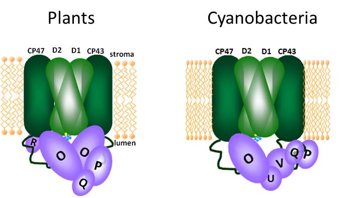
Results: The PPD1 RNAi mutants with the lowest PPD1 expression were extremely small and pale green plants. Analysis of chlorophyll fluorescence showed that the mutants had much higher levels of fluorescence, indicative of an over-reduced plastoquinone pool and problems on the PSI-side of the photosynthetic electron transfer chain. Specific measurements of PSI activity showed that the PPD1 RNAi mutants had reduced amounts of active PSI reaction centers. However, energy could be transferred to these reaction centers by an alternative antenna system (LHCII). Moreover, the function of the PSI centers which did accumulate was not normal. Further analysis of protein accumulation in the thylakoids of the PPD1 RNAi mutants revealed there were specific problems in the accumulation of proteins on the lumenal side of PSI. In wild-type plants, the PPD1 protein was found to be associated with a thylakoid protein complex of ~300 kDa, which is smaller than any PSI complex.

2D gels showing thylakoid protein complexes in WT and PPD1 RNAi mutant. 1,2,4 complexes are forms of PSI; 3 is ATP synthase

2D gels showing thylakoid protein complexes in WT and PPD1 RNAi mutant. 1,2,4 complexes are forms of PSI; 3 is ATP synthase

Conclusions: The PPD1 functions in the proper assembly of PSI components on the lumenal side of the complex. In this area, PSI contains an extrinsically associated protein, PsaN, as well as extensive loop regions of the membrane proteins PsaA, PsaB and PsaF. All of these components create the binding environment for the soluble electron carrier, plastocyanin, which delivers electrons from upstream in the transfer chain. Reduced amounts of PPD1 affect the accumulation and assembly of these components. The mutant plants try to compensate for this loss of functional PSI by shifting some of the LHCII antenna such that it can funnel energy into PSI (the default for LHCII is to drive energy into PSII). The PPD1 protein is not considered a subunit because it was not found to be associated with fully assembled PSI complexes, but a smaller protein complex.

Think Ahead: The assembly of PSI is not a well-characterized biological process because, unlike PSII, PSI is an extremely stable enzyme. Thus, once it is assembled, the complexes can function properly for very long periods of time. Because it is a rare process, it is difficult to study. The original characterizations of PSI subunit mutants were performed many years ago, and it may be interesting to give them a fresh look with respect to PPD1 and some of the antenna effects we described. Identification of the other proteins in the complex with PPD1 (other PSI subunits perhaps) may help to define a PSI assembly intermediate. The secondary effect that the PPD1 mutation had on the antenna system will also be interesting to follow-up on because we don’t really know all of the details governing how plants allocate light energy between the photosystems. It is a sophisticated system with multiple layers of control.


Tale 2

“Nothing that we do, is done in vain. I believe, with all my soul, that we shall see triumph.” Charles Dickens

While scientific endeavors may have their dark moments, scientists tend to think that ultimately their research will see triumph. In the world of academia, this means publication in a peer-reviewed journal. Thus, all of the experiments that were done leading up to that publication but not included in it are not done in vain. They helped to work out the procedures necessary for acquiring the data that did appear in the figures. They were experiments that yielded negative data which eliminated hypotheses. Alas, those are never published.* It may be useful for scientists to know what wasn’t, but publishers only want to tell the stories of what was. (Hey, that almost sounds like Dickens too.)

The PPD1 story started with a blanket search for functions of the PPD family of proteins in the thylakoid lumen. They must be doing something to help plants photosynthesize, right? I was hopeful that maybe one of them had something to do with my favorite enzyme PSII. The way I chose to attack this problem was to characterize mutants of each of these proteins in Arabidopsis.

The easiest way to acquire Arabidopsis mutants is to order T-DNA lines (insertion mutants) for your gene of interest from the ABRC stock center. They send you seeds and you check to see if the mutants are useful or show a phenotype. There were two available lines (independent insertions) for the PPD1 gene and both of them were less than helpful to me. One line that another postdoc had been working with that had been passed on to me, which may have shown a phenotype, turned out to be heterozygous (not a complete mutant; still contains one wild-type PPD1 gene and should be normal). I couldn’t replicate any subtle phenotype, but neither could I find any homozygous (complete mutant) plants. Ever. I spent a lot of time verifying that that particular T-DNA mutant was embryonic lethal for homozygotes. Strange, but possibly extremely interesting. However, before taking this as a fact, it had to be confirmed that the homozygous lethal phenotype was because of the mutation in the PPD1 gene and not some other random mutation elsewhere in the genome. This can be sorted out by backcrossing heterozygote mutants to wild-type plants a few times and trying to recover the mutants. Ultimately, my experiments showed that the link between the PPD1 mutation and the embryonic lethal phenotype was not so absolute. At the same time, I was growing the other T-DNA mutant and it was proving to be equally unstable. Some plants would show a variegated phenotype, streaked leaves with patches of green and pale yellow. Other plants looked normal. I could never consistently link the phenotype to the ppd1 mutant genotype. With all of these inconsistencies in results, I decided that the T-DNA mutants were not useful in telling me anything about PPD1 function.

The alternative approach to Arabidopsis mutants is to use the RNAi technique to selectively suppress the expression of your favorite gene. While I was wrestling with the PPD1 T-DNA lines, I began the process of generating my own PPD1 RNAi lines. These plants turned out to be the most useful for figuring out what PPD1 does. When screening through these mutant individuals, there was a range in what the individual plants looked like- some looked almost normal while others were very small and pale. This is the good thing about RNAi because this range among individuals gives so a sort of picture of what is happening when the expression of the gene of interest is turned up or down over a gradient, like tuning up a brightness or volume knob on an old TV.

There was very little material to work with for the most severely affected PPD1 RNAi plants, but the biophysical measurements I could do on the tiny leaves indicated there was no problem with PSII. The defect was further downstream, probably in PSI. When using an instrument to specifically measure PSI function it was clear that was where the problem was. I would have to learn more about the PSI complex to say enough to turn my results into a publication, but at least I knew where this was going.


“There is prodigious strength in sorrow and despair.” Charles Dickens

The same week as my PSI results, I received an after-hours e-mail from my PI with the link to the following journal article: PsbP-domain protein1, a nuclear-encoded thylakoid lumenal protein, is essential for photosystem I assembly in Arabidopsis, Liu et al 2012 Plant Cell. When I quickly skimmed the abstract, my heart sank. My response was $%&^!, $%^@!, #$%@!, *&$%! I think I even drowned my sorrow in a pint of Hagen-Daz. There was only the slightest glimmer of satisfaction from the validation that researchers on the other side of the globe had come to the same conclusion as me.

Validation is not the name of the game. You see there is no prize for second place in scientific publishing. When you are the first group to publish a new idea, you have more control over the limits of the tale are. When you are second place, you cannot merely confirm what has been done (PPD1 has something to do with PSI). You must take it further, press on to unravel more details. Pressing on into the details of PSI territory was not really what I wanted to do.

However, after carefully reading what Liu et al had done, I reassessed my data and found a way to move forward. They had managed to characterize a clean T-DNA line, and the homozygous mutant plants they worked with were completely devoid of PSI. The small pale plants had to be grown on sucrose-containing medium since they could not support themselves photosynthetically. In my work, the RNAi lines allowed me to characterize plants that were very sick, but could still grow on soil. They accumulated some PSI, which could be analyzed more closely. Of course, that meant that I had to do a lot of experiments on precious little material. These experiments meant using a lot of brute strength just to get enough material for the experiments (spectroscopy measurements and blots, oh the blots!), investing time in fine-tuning protocols and money in antibodies for our second-favorite thylakoid complex.

“Vengeance and retribution require a long time; it is the rule.” Charles Dickens

Pushing forward with the experiments was difficult and took quite a bit of time. The sickest of the PPD1 RNAi lines were very small and would not set seed. Getting enough material meant screening for primary transformants every time. Learning the literature for a different enzyme complex was challenging. The papers describing the original characterizations of PSI subunit mutants were at least a decade old and often lacked data I would have liked to have seen. Not really flaws with that work; it’s just that what was not necessary for that work would have been extremely helpful to me.

Eventually, all of my data was written up in manuscript form and submitted away for peer review. I dabbled in other projects waiting the weeks until reviews came back. It took longer than usual, which meant only one thing- it was sent to a third reviewer. Yes, the two reviewers that initially evaluated my work had such differing views as to what my manuscript’s fate should be, a third reviewer was enlisted to help the editor in making the appropriate decision. Please revise with additional experimentation and there were specific concerns about how we went about doing some of our experiments.

Yes, a long time is the rule. I spent the next months painstakingly addressing the reviewer’s points with new experiments. One issue was how we estimated the amount of PPD1 protein in the mutants. With our antibody and a number of variations of our gel system, the PPD1 protein ran at the same molecular weight as the LHC proteins- the most abundant membrane protein on earth. These proteins obscured the signal for PPD1 such that we could never reliably estimate its amounts on denaturing gels. It either could not be seen or samples would require too much handling and treatment to consistently give a signal. However, PPD1 could be perfectly detected on native gels because the LHCs were nowhere near it in that system. Finally, I had point-by-point addressed all of the issues, revised the manuscript and created new figures.

We were ready to try again, but the tone of one of the reviewer’s comments gave us pause about resubmitting. Sure we had responses, but the original comments seemed like they would never invite satisfaction. There were some things about our results that would just not change. Experiments were done properly and yes, the results were still slightly unexpected. We would not be making up data because it would be easier for reviewers to accept. That is a cardinal sin in science, and a separate rule that should never be broken. For the revised manuscript, we took the chance on submitting to a separate journal with different reviewers for the chance of a favorable decision. This wager did not pay off because the two new reviewers had a completely separate set of comments to be addressed, many of which seemed impossible to satisfy with our sample limitations. We declined the invitation to revise and resubmit and ultimately resubmitted to the original journal. It went back to the original reviewers who seemed mostly satisfied with the improvements. Of course, there were some new comments by the reviewers that we could just not accommodate; not because the concerns were invalid, but because the questions went well beyond the scope of our work. There will always be more experiments to be done, but we firmly and politely stated we would not be addressing the new questions our latest experimental results sparked. We could only speculate as to future possibilities in the discussion section.

“A multitude of people and yet solitude.” Charles Dickens

In the case of this particular project, it was a multitude of data, yet not figures. I have numerous notebooks filled with raw data from experiments related to PPD1. In science, you go through a lot of preliminary work to get the answer, but when you show your work it must be much neater (certain colors, intensities, certain samples in certain orders). It’s like taking a math test and having a separate scratch paper (the lab notebook), but your answer is only a single circled number or neat graph (the manuscript figures). In this particular case, it was a multitude of blots. I cannot tell you how many blots I developed for this project. I practically lived in the dark room for months, eagerly waiting for films to emerge from the developer, praying the signals would be beautiful enough for figures.**

“I wish you to know that you have been the last dream of my soul.” Charles Dickens

Appeasing the reviewers in this final round felt a lot like the emotions in this quote. I had started forming the publication framework haphazardly because it wasn’t on a topic that I found exciting. Admittedly, I was only trying to do just enough to get acceptance. Even though my sentiment for some of my reviewers was more akin to a different saga, their requests did make the story better and forced me to expand my general knowledge on PSI and technical expertise in new protocols. All research can continue ever and on, but lines must be drawn somewhere because of the universal limits of effort, time and finances. I felt that the story was finally new and good with enough potential tangents to drive future research by possibly myself and others in the field. I had finally come to the point that I didn’t just want it done for the sake of adding another publication to the tally, but I wanted it published because the results deserved to be part of our body of photosynthesis knowledge.***

“It is a far, far better thing that I do, than I have ever done; it is a far, far better rest that I go to than I have ever known.” Charles Dickens

My PPD1 manuscript was eventually accepted at the Journal of Biological Chemistry this summer after a very long road of experimental struggles and research-related drama. Of all of my publications, this was definitely the most difficult to get to the point of publication. It is probably at the bottom of the list of my works if I had to rank them my favoritism based on any scale. This post was actually quite difficult to write; I so long to leave it in the past. However, I can now recognize that it is one of the better things I have done just to not give up on it. And after all the co-authors thought that everyone who would ever be interested in the PPD1 protein would have written the paper or reviewed it, we get an e-mail from another research group requesting our PPD1 antibody because their work may have a link to PPD1. “We read your paper with great interest,” they said in an e-mail sent a mere two days after our accepted manuscript appeared on-line. “They did!” I laughed. I sent them a sample, some behind-the-science instructions and well wishes. Apparently, my perseverance wasn’t just a better thing in terms of racking up publication numbers on my CV, but also for some other researchers embroiled in their own scientific epic. The best of times and the worst of times indeed.

As for me, it is a far, far, better rest that I go to than I have ever known as well. It’s not just a project change and definitely not the guillotine. Announcement coming soon to the blog.


Johnna

*However, Elsevier is launching a new journal where you can publish those results. Introducing the new journal New Negatives in Plant Science.

**There is a popular song this summer by Lil Jon and LMFAO “Shots”. There are not many lyrics. The rapper mostly just says shots over and over the backbeat track. To stave off insanity, or perhaps the opposite, I would sing my own version of the song “Blots.” All my biochemists, where y’at? Let’s go. When I walk in the lab, gloves on me, with the antibodies, I love chemiluminescence, I came to develop, lights off, it’s on! Blots, blots, blots, blots, blots, blots, blots, blots, blots, PPD1, blots, blots, blots, blots, blots, PsaB, blots, blots, blots, blots, blots, blots, LDS-PAGE, blots, blots, blots, blots, blots, Blue native, blots, blots, blots, blots, blots… If I don’t do these blots, I can’t resubmit! You get the idea. For visual effect, you can also picture me making it rain with x-ray films. Hey, what do you know another parody for the blog.

***Although if it would not have been accepted, I had threatened to just dump all of my results on this blog anyway and be done with it. Not sure how it ranks in terms of impact factor though.

References and Links:

http://www.jbc.org/content/early/2014/07/09/jbc.M114.589085

https://www.goodreads.com/work/quotes/2956372-a-tale-of-two-cities

Dangerous Photosynthesizers

Because photosynthetic organisms are the energetic foundation of our biosphere, we always tend to think of them as allies, organisms with a positive connotation. Their trademark color green is universally linked with goodness, growth, and life. However, there are some bad apples in the bunch that just seem to have it out for us heterotrophs. Well, maybe not apples (though I’m sure there are some poisonous apples out there somewhere*), but nature is filled with examples of poisonous plants. Many toxins and pharmaceuticals have botanical origins.

The focus of today’s blog post sinks even lower- algae, pond scum, cyanobacteria. Most of these aquatic photosynthesizers quietly convert sunlight to biochemical energy without any ill effects to anyone. I’ve written previously, that under the right conditions (warm and nutrient-rich waters) these otherwise inconspicuous organisms bloom in great numbers and overwhelm their environments. Like all life on earth, algae are programmed to capitalize on favorable conditions for reproduction. The ultimate crashes of these blooms can result in aquatic dead zones, areas with dissolved oxygen levels too low to support life.**

Algal bloom in Lake Erie 2011 from the NASA Earth Observatory Credit: Jesse Allen and Robert Simmon via Wikimedia

However, in some cases, the effects of these algal ‘blooms’ go beyond sheer numbers. Some algae produce toxins which cause serious health problems for those of us heterotrophs sharing their environment. The individual constituents of these ‘harmful algal blooms’ (HABs) measure in at ~1-2 µm, but they can wreak all kinds of havoc on the scale of large cities. HABs can stop your summer fun by forcing beaches to close or eliminating certain shellfish from your diet, but this summer the problems went beyond recreation to something much more fundamental- potable water.

Clean, safe drinking water is a fundamental service of human civilization. In our modern society, just turn on the tap, cook, clean, bathe, drink. It has been such a staple of American cities that it is taken for granted. That is, until it’s no longer available. That was the exact situation in Toledo earlier this month. A large American city, in the year 2014, was without safe drinking water for a whole weekend. Approximately, 500,000 citizens were affected. All because of toxic algae.

Microcystin-LR chemical structure Credit: cacycle via Wikimedia

In this case, the culprit was a bloom of cyanobacteria which produce the toxin microcystin. This molecule is harmful to the health of humans, pets and wildlife by acting as a liver toxin. It also has neurotoxic effects. The toxicity of microcystins has been extensively characterized and long-known to be associated with certain cyanobacterial species. Because of the potential adverse effects of microcystin-producing cyanobacteria on modern water supplies, treatment facilities routinely check the levels of this toxin. Only one part per billion of this molecule is considered acceptable. When a microcystin-producing algal bloom occurs near the intake of a municipal water supply (as it did for Toledo this month) the facilities can quickly be overwhelmed causing the water supply to exceed acceptable microcystin levels. The situation is compounded by the fact that microcystins are resistant to boiling. While boiling water may destroy other toxins or contaminating bacteria, it only concentrates microcystins. In order to bring the toxin levels down, the problem must be addressed at the water treatment facility (using methods like activated carbon, ozone treatment and membrane filtration). By adjusting the normal procedures to account for increased microcystins, the water supply can be treated to once again safe levels. All of this is accompanied by exhaustive analysis of microcystin levels and vigilant monitoring after the incident.

Scientists and government agencies are always working to monitor our water systems for HABs. Check out some of the links below for descriptions of continuous efforts to monitor our environment for HABs. How do we get to the point of 500,000 people without water for a weekend with everyone watching out for it? I mean, we’re watching it from space! Even with all of these sophisticated tools and models, nature can be surprisingly swift. Check out these images and reports from the NOAA Great Lakes Environmental Research Lab on Aug 1, 2014 and Aug 4, 2014 showing false-colored images tracking the algal growth.

Simultaneously in the news cycle with the Toledo water crisis, two Ebola patients were being treated in premier isolation facilities on American soil. The nation’s attention was rapt with the details of their treatment and speculation was rampant as to the possibility of an outbreak in America (an infinitesimally small probability not worth talking seriously about among scientists and epidemiological experts, but a great ratings-driver). Worldwide, the Ebola death-toll numbers in the thousands, not just from this year, but ever. Based on pure body count, there are many deadlier infectious diseases, which we as the public dismiss more easily. Beyond those numbers, the lack of clean water for drinking, food preparation and sanitation results in the deaths of ~3 million people every year across the globe. A safe and reliable water supply, as a basic right, continues to elude human civilization.

Water, water everywhere, but not a drop to drink… Credit: de:Benutzer:Alex Anlicker

HABs are only a part of the world’s water problem. However, the disruption of Toledo’s water supply should have been an event that caught our attention and held it for a while longer. It may be easy to turn on the tap, but getting the clean water to that point takes a significant amount of effort with infrastructure maintenance, monitoring and treatment. All of these things are largely invisible to us in modern society. Unfortunately, all of these things are affected by other societal choices like economics, aesthetics, environmental regulations, and the practices of our agricultural systems and other industries. As a society, we should start having the longer, difficult conversations necessary to attack this complicated problem rather than the transient chats that occur when we are in crisis mode. Find out about your community’s water situation and the issues related to your supply. Talk to your community leaders today to ensure that safe water is part of your future.


Johnna

*Hey, it’s hard to transition away from the summer’s Disney theme completely in a single post. However, I’m not just talking about the evil queen’s poisoned apple from the fairy tale. Apples have a huge amount of genetic diversity and I’m sure there are some varieties out there that are poisonous or so foul-tasting that you would think they are. After all, apples concentrate toxic substances in their seeds.

http://www.thepoisongarden.co.uk/atoz/malus_john_downie.htm

**In case you’re wondering, this year’s Gulf of Mexico hypoxic zone measured ~5500 square miles. That’s not breaking any records for size, but still about as large as the state of Connecticut. Read more about it here.


References and Links:

http://oceanservice.noaa.gov/hazards/hab/

http://www.cop.noaa.gov/stressors/extremeevents/hab/current/noaaHab.aspx

http://www.cop.noaa.gov/stressors/extremeevents/hab/current/HAB_Econ.aspx

http://www.cdc.gov/nceh/hsb/hab/default.htm

http://www.whoi.edu/redtide/page.do?pid=14898

http://www2.epa.gov/nutrientpollution/harmful-algal-blooms

http://www2.epa.gov/nutrientpollution/effects-human-health

http://www.dispatch.com/content/stories/local/2014/08/04/this-bloom-is-in-bad-location.html

http://www.lakeeriewaterkeeper.org/

http://www.gulfhypoxia.net/news/default.asp?XMLFilename=201408111452.xml

http://en.wikipedia.org/wiki/Microcystin

http://iaspub.epa.gov/tdb/pages/contaminant/contaminantOverview.do?contaminantId=-1336577584

http://www.weather.com/health/what-you-need-know-about-microcystin-toledos-water-toxin-20140804

http://water.org/water-crisis/water-facts/water/

http://water.org/water-crisis/one-billion-affected/

http://www.cdc.gov/healthywater/global/wash_statistics.html

PSII is a Fixer-Upper

Frozen

The final song from our Frozen parody reminds us that just because something is damaged or broken, doesn’t mean it can’t become whole again with a few repairs. This sentiment makes it the perfect theme song for my favorite enzyme, Photosystem II (PSII). Here’s the Disney version…


Is it the slower QB reduction?

Or lack of water oxidation?

No use trying state transitions,

Although we know it transfers well, PSII ends up photodamaged

Such photoinhibition is such an imposition

So it’s a bit of a fixer upper

So it falls behind

Its peculiar mechanism

Is a one-of-a-kind

So it’s a bit of a fixer upper, but we’re certain of this one

You can fix this fixer upper with a newly made D1

Is it the oxygen singlets?

In chlorophyll protein ringlets?

Is it the way the water splits?

Electrons zipping towards quinones

Causing D1’s aches and groans?

Now permanently on the fritz.

PSII’s just a fixer upper

It needs a protein exchange

Its phosphorylation is confirmation

That something is strange

So it’s a bit of a fixer upper

Plant cells know what to do

The way to fix this fixer upper

Is to make D1 anew

Damaged PSII is a bit of a fixer upper

That’s a minor thing

Just disassemble then reassemble

Voila just like recycling!

So PSII’s a fixer upper

It’s function is nixed

Get the damaged protein out of the way

And the whole thing will be fixed

We aren’t saying to remake the whole thing

‘Cause that’s just too much work

We’re only saying that light’s a force that drives PSII berserk

Electrons just excite the wrong things if they go off their normal path

Changing out the D1 protein erases all their wrath

New D1 clears the path!

All PSII’s are fixer uppers

They’re made to fizzle out

DegP, phosphorylation

FtsH, degradation

Get them on the repair route

All PSII’s are fixer uppers

When the electrons start to move

The only fixer upper fixer

That can fix a fixer upper is

To get the damage removed


Photosystem II performs the unique reaction of splitting water to form oxygen and extract electrons used to fuel photosynthesis. Not all of this energy goes in the direction that it should. When energy gets backed up in the system or electrons venture off of the designated path, irreversible damage to the proteins can occur. This damage means that PSII doesn’t work anymore. Because this photodamage is an unavoidable hazard, photosynthetic organisms have an efficient way of dealing with this problem.

For one thing, the D1 protein at the heart of the PSII complex bears the brunt of the irreversible damage. This makes sense because the D1 protein also coordinates many of the cofactors that comprise the electron transfer pathway through the system. On the one hand, damage to this one protein means function gets knocked out as well; on the other hand, it means that the damage is concentrated on just one protein. So, to fix it and restore function to the complex means photosynthesizers mainly focus on replacing one protein, not twenty. That’s what has to happen. The damaged protein must be replaced by a newly synthesized copy.

It sounds simple enough, but anyone who’s done any fixer-upper work knows there’s more to it than that. Repairing the damage starts with recognition; there must be systems in place to differentiate functional PSII from damaged PSII. Phosphorylation of certain residues on PSII subunits labels those complexes as targets for repair. These labels are interpreted by specific proteases, which then remove and chop up the damaged D1 protein. Next, a newly made D1 protein is inserted into the complex to restore function.

The proteases involved in removal of the damaged D1 protein are DegP and FtsH. Researchers still debate over which one is more important, but it is likely to be a combined effort by both. Also, because the D1 protein is located within the middle of the PSII complex, many other subunits and cofactors must partially or transiently disassemble as a result of D1 protein removal. Exactly how this works and what additional proteins are involved in this process are active areas of research in the photosynthesis community.

Photosynthesizers don’t give up on their PSII complexes just because they get a few dings. The constant recycling of PSII complexes through this repair process ensures that the light reactions of photosynthesis will continue to churn away, even in bright light (more energy, higher rates of damage). It may seem like a lot of trouble to run this elaborate repair shop, but it’s still easier than starting from scratch each time PSII is damaged.


Johnna


References and Links:

http://www.disneyclips.com/lyrics/frozenlyrics9.html

http://en.wikipedia.org/wiki/Photoinhibition

PSII damage and repair

http://www.ncbi.nlm.nih.gov/pmc/articles/PMC2889791/

http://www.ncbi.nlm.nih.gov/pubmed/10322546

http://www.pnas.org/content/91/15/7222.full.pdf