Tag Archives: Rubisco

The Dark Side Comes to Life in Fluorescent Colors

Something new under the sun… carboxysome assembly in real time.

Today’s post features research out of the Kerfeld Lab from a close colleague of mine, Jeff Cameron.* After doing his thesis work on redox homeostasis in cyanobacteria, he moved on to a postdoc on the dark side of photosynthesis.** His new paper just out in Cell today sheds new (fluorescent) lights on a longstanding question in cyanobacterial carbon fixation.

Cameron et al. Cell Volume 155, Issue 5 2013 1131 – 1140

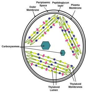
Schematic for cyanobacterial cell structure showing membrane systems and carboxysomes.

Schematic for cyanobacterial cell structure showing membrane systems and carboxysomes.

I’ve mentioned before that of the oxygenic photosynthetic organisms, cyanobacteria have an elaborate system for concentrating CO2 at the site of Rubsico. This prevents Rubisco from getting confused and using O2 instead, a costly mistake for the cells. Sure, these cells have an array of transporters as well as carbonic anhydrases to interconvert CO2 and bicarbonate. But the real heart of the cyanobacterial Carbon concentrating mechanism (CCM) is the carboxysome- an intracellular microcompartment where the cells corral all of their Rubsico. This structure allows the cells to handily funnel all of their acquired CO2 so that it can be fixed by Rubisco. No, I didn’t say it’s an organelle. Cyanobacteria are prokaryotes; while they may break the rules by having an internal thylakoid membrane system, they do not have true organelles in the sense that eukaryotes do.

So what are they exactly? They are protein-based structures, in which an array of Rubsico is encapsulated by hexameric and pentameric shell proteins. Carboxysomes have been well-studied from a structural standpoint and the major protein components have been identified.

Schematic model of the α-carboxysome assembly containing RuBisCO small (dark green) and large (green) subunits and carbonic anhydrase (red). The shell is composed of hexamers (blue), pseudohexamers (light blue, magenta, and light green), and pentamers (yellow) From Kinney et al 2011 Photosynthesis Research

However, all of this beautiful structure work doesn’t provide any information on the dynamics of these components as the cells must construct them.

Cameron and co-authors describe an experimental system that allowed them to follow the assembly of carboxysomes from scratch. This system also allowed them to eliminate carboxysome subunits one at a time and monitor the effects on assembly. From all of these results, they were able to piece together a model for carboxysome biogenesis, something that wasn’t possible from looking at static pictures from single mutants.
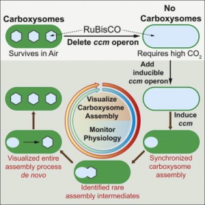
Graphical Abstract Cameron et al 2013 Cell Biogenesis of a Bacterial Organelle: The Carboxysome Assembly Pathway

Graphical Abstract
Cameron et al 2013 Cell
Biogenesis of a Bacterial Organelle: The Carboxysome Assembly Pathway

Here’s how the method breaks down:

Observations: Mutants of Synechococcus elongatus PCC 7942 that lack the carboxysome structural proteins cannot confine their Rubisco to small areas and require higher than air-levels of CO2 to grow photosynthetically. Individual mutants of the various carboxysome subunits have variable effects on carboxysome structure, but these are not useful for understanding how the carboxysome subunits come together in a temporal sequence during assembly.

Hypothesis: If a system could be generated to synchronize carboxysome assembly (going from no carboxysomes present to fully assembled carboxysomes), then researchers could probe the effects of the presence/absence of structural components on assembly. This information will allow researchers to deduce the order of assembly events.

Experiment: A synchronized carboxysome assembly system was generated by starting from a mutant of Synechococcus elongatus PCC 7942 lacking all carboxysome structural subunits (no carboxysomes). These structural subunits were added back to this mutant background (as a complete set and as with individual missing subunits) under the control of an inducible promoter. These various cell lines were also engineered to express a fluorescent version of Rubisco were produced to serve as a marker for carboxysome assembly (diffuse fluorescence = no assembly; tight fluorescent spots = assembly).

Results: The control cell line (fluorescent Rubisco + complete set of structural carboxysome subunits under inducible control) showed a diffuse localization of Rubisco throughout the cells prior to the induction of carboxysome genes. After the addition of the inducer chemical, the fluorescence from Rubisco focuses into small green spots indicative of carboxysome assembly. The cases where less-than-the-complete-set of carboxysome subunits were added back to the original mutant cell line, the induction yielded variable results for carboxysome assembly. Some did not assemble any carboxysomes (diffuse Rubisco fluorescence) while others showed intermediate patterns of Rubisco fluorescence (abnormal or partially assembled carboxysomes).

Conclusions: The authors deduced a new model for carboxysome assembly. First Rubisco proteins and the subunit CcmM aggregate together to for pro-carboxysomes (PCs). CcmN is then added to the PCs and begins to associate with the outer shell proteins CcmK2 and CcmO. Finally, when the CcmL protein is incorporated, the carboxysome completely enclosed by its protein shell and released from the PC.

Think Ahead: Carboxysomes are a prototype for other bacterial microcompartments. Other bacteria use similar protein-based microcompartments for specialized metabolism. This system and the insights on the timing sequence of its assembly can applied to these other systems. In any case, these experiments lay the groundwork for engineering similar microcompartments for medical or biotech purposes. For instance, plants might be coerced to make carboxysomes for more efficient carbon fixation. Alternatively, it may be possible to create novel microcompartments in other bacteria for the purposes of efficiently making useful metabolites.

Check out the link below for the fluorescence microscopy video of carboxysome biogenesis. Sorry, there’s a paywall for 12 months.

http://www.sciencedirect.com/science/MiamiMultiMediaURL/1-s2.0-S0092867413013627/1-s2.0-S0092867413013627-mmc3.mp4/272196/FULL/S0092867413013627/fe4fe2db6252ffa45cbd984455447380/mmc3.mp4

UPDATE: There is a free version of Supplemental Video #2. Thanks JGI!

This is after all carboxysome genes have been turned on and you can watch them move and even see some bud off after awhile. (I still prefer the other video where you can watch them form from nothing.) Also, in the current edition of the video caption, they credit James Cameron instead of Jeff Cameron. Sure, James does great cinematic work- just not this one. (It should be fixed on Monday.)

Jeff Cameron, the James Cameron of carboxysome cinema.


Johnna

*Jeff and I were graduate students together in Himadri Pakrasi’s lab at WashU. I think he still hears my voice in his head when he does biochemical purifications. “Fast and Cold! Jeff. Fast. And. Cold.”

**Yes, yes, I know. Light-independent. Whatever. ‘Dark’ makes for catchier writing and storytelling.

References:

http://www.sciencedirect.com/science/article/pii/S0092867413013627

http://www.microbemagazine.org/index.php?option=com_content&view=article&id=1863:the-carboxysome-and-other-bacterial-microcompartments&catid=464&Itemid=826

http://link.springer.com/article/10.1007%2Fs11120-011-9624-6/fulltext.html
Better photosynthesis in rice from an unexpected source

Something new under the sun… A gene for better photosynthetic rate in rice.

It’s something we knew before, but not in relation to photosynthesis. It’s also something breeders selected for without knowing the actual mechanism. Now it’s a new piece of the puzzle for increasing rice yields and meeting our 2050 yield goals. It’s in this recent report:

A natural variant of NAL1, selected in high-yield rice breeding programs, pleiotropically increases photosynthesis rate

Takai T, Adachi S, Taguchi-Shiobara F, Sanoh-Arai Y, Iwasawa N, Yoshinaga S, Hirose S, Taniguchi Y, Yamanouchi U, Wu J, Matsumoto T, Sugimoto K, Kondo K, Ikka T, Ando T, Kono I, Ito S, Shomura A, Ookawa T, Hirasawa T, Yano M, Kondo M, Yamamoto T.

Sci Rep. 2013; 3: 2149

The different aspects of photosynthesis play a major role in determining the yields of staple crops like rice. There must be a balance among sink size (the amount of sugars stored from photosynthesis), source strength (the capacity to make sugar from photosynthesis), and carbohydrate translocation (the movement of sugars from metabolism to storage). Natural rice cultivars have a wide range of photosynthetic rates per given CO2 levels. This rate is governed by how efficient plants are at taking in CO2 from the atmosphere and how fast CO2 is processed by Rubisco. This physiology is well-characterized but the genes underlying the complex trait of enhanced photosynthesis remain elusive. Takai and co-authors have identified a locus (section of DNA) responsible for photosynthesis rate by controlling CO2 carboxylation. Here’s how they did it.

Observations: Naturally-occurring rice varieties have a range of photosynthetic rates and their genomes should contain useful gene variations for this complex trait.

Hypothesis: The high-yielding, high-photosynthesis rate rice varieties contain specific genes underlying these traits. Identifying these genes will identify new targets for breeding programs aimed at developing rice varieties with even higher yields.

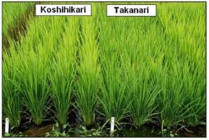
Experiment: Rice lines were chosen for genetic analysis to identify the genes responsible for controlling photosynthesis rate. One line (Takanari) represented the highest-photosynthesis rate of any cultivated rice variety. The other (Koshihikari) represented the leading, high-yielding rice variety with the greatest difference in photosynthetic rate relative to Takanari. Genetic mapping techniques were used to identify the specific gene responsible for the high rate of photosynthesis in the Takanari variety. Physiological measurements were also performed to figure out why this gene confers better photosynthetic rates.

Takai et al Fig 1B Sci Rep. 2013; 3: 2149 Creative Commons License

Takai et al Fig 1B, Sci Rep. 2013; 3: 2149
Creative Commons License

Results: One locus named GREEN FOR PHOTOYSYNTHESIS (GPS) was identified as a contributing factor for increased photosynthetic rate. Takai and co-authors determined that the better photosynthetic rates were due to an increase in Rubisco content, which increased the rate at which CO2 could be converted into sugars. When the researchers identified the DNA sequence of the GPS locus, they found it was a gene that had been previously characterized and named NARROW LEAF1 (NAL1). NAL1 is a plant-specific protein important for plant hormone* transport, which controls plant growth. It had been previously established that nal1 mutants resulted in dwarfed rice plants, but photosynthesis rates had not been examined. So, the researchers in this study went back and had a look at photosynthetic rates in previously characterized nal1 mutants. Sure enough, they had higher photosynthetic rates too. The specific GPS sequence variants identified in this study, however, resulted in higher photosynthetic capacity without the dwarf plant trait. This is because of the difference in severity of the GPS and nal1 mutants; previously characterized nal1 mutants were complete loss-of-function mutations, while the GPS variant in this study was still partially functional. When the researchers looked at the leaves of the GPS variants, they found increased cells and thicker leaves. This is consistent with an abnormality in the function of a gene controlling plant hormone function, and the photosynthetic increase is a secondary effect.

Conclusions: The disruption of the GPS(NAL1) gene in rice results in higher photosynthetic rates because of better CO2 fixation. This trait is intertwined with control of plant stature via a plant hormone pathway. So, leaf shape at the cellular level can have a profound effect on how efficient photosynthesis is. With the identification of this gene, the scientists were able to go back and look at more rice varieties and check for GPS. As it turns out, even though breeders didn’t know the molecular details behind it, this trait had been selected for in high-yielding rice breeding programs.

Think Ahead: Using the GPS(NAL1) gene as a marker for photosynthetic productivity can improve breeding strategies for making higher-yielding rice varieties. This study demonstrates that scientists must look beyond the genes involved in the biochemical reactions of photosynthesis to improve photosynthetic rates. In this case, a gene controlling leaf cell structure was a major determinant for increases in CO2 fixation. While this link is clear, we have no idea what the precise function of the GPS(NAL1) gene is. The story gets more complicated when trying to translate these findings into actual rice grain yield increases. The GPS(NAL1) is not the whole story because the presence of this gene variant alone is insufficient to give appreciable grain yields without the presence of other genes controlling yield or the addition of nitrogen fertilizer. This report provides great new information and tells us we still have more to do on the way for optimizing rice yields.

Check out the full article here**: http://www.ncbi.nlm.nih.gov/pmc/articles/PMC3756344/

This work represents a huge effort in plant genetics just to identify GPS. These researchers used a technique called quantitative trait loci (QTL) mapping to sort through which section of the rice genome was responsible for the trait or phenotype they were interested in. Let’s just say it involved growing a lot of individual rice plants, analyzing their physiology and using statistics to separate what was important from what was insignificant. Because the rice genome is completely sequenced, these researchers were able to determine the specific gene responsible for the trait they observed. They could directly compare the standard sequence to the sequence of their high-photosynthesis rate variety and leverage this information into more specific hypotheses for underlying function. This type of work again illustrates the confluence of specialties (genetics, statistics, physiology) required to answer complex biological problems. Scientists have to be willing to follow the results into whatever field it takes them- especially when the path wanders from photosynthetic carbon fixation to hormones that control plant development and leaf structure.

Johnna

* Yes, in case you didn’t know, plants make hormones too. While they are chemically quite distinct from the animal hormones you may be familiar with (adrenaline, testosterone, estrogen etc.), they perform analogous functions to control things like plant growth and other physiological responses. Plant hormones are a difficult topic because of their diverse functions and complicated interactions.

** Open access!!!